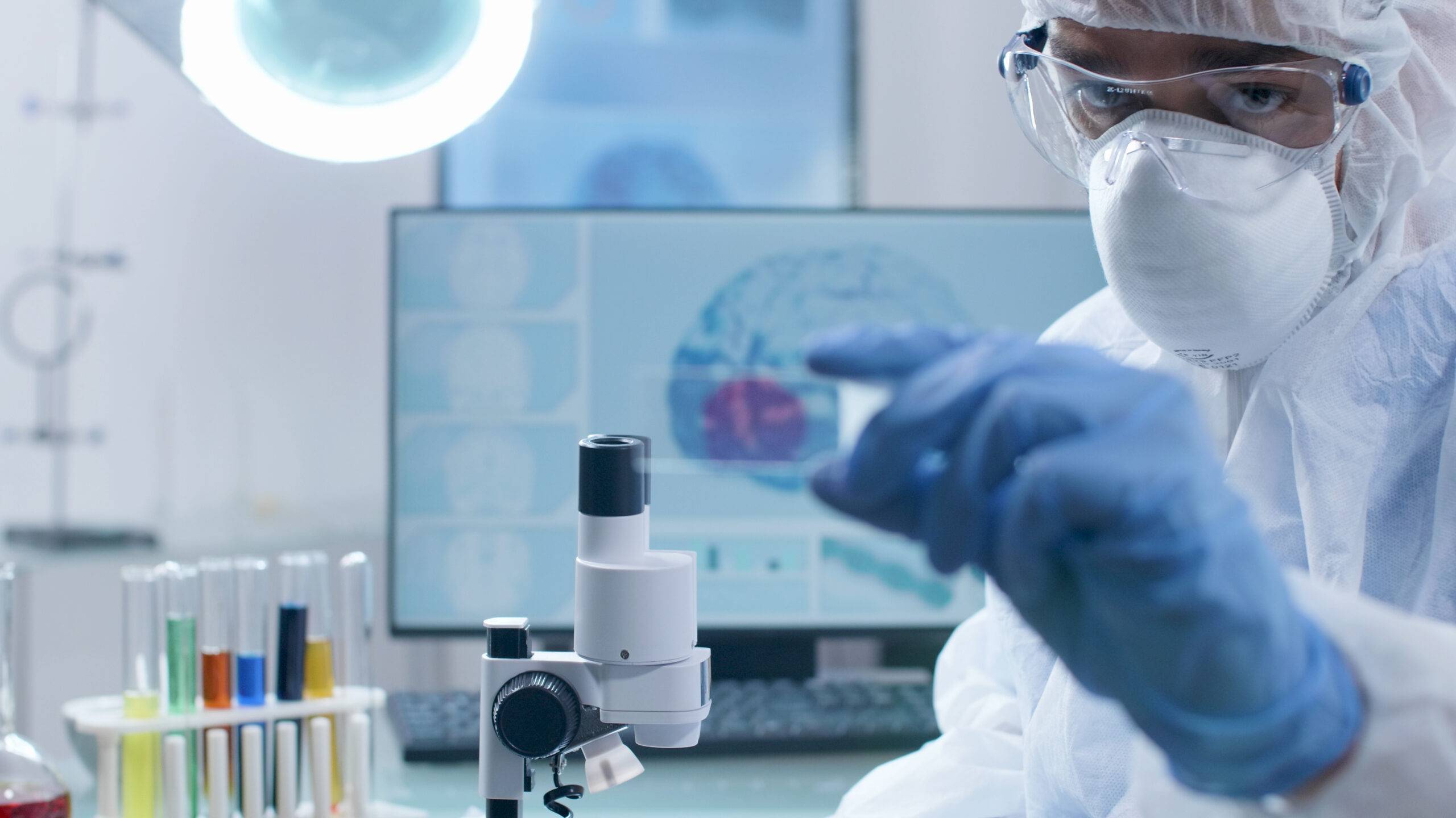
In order to discover and define the most basic primary and secondary endpoints in clinical trials, the process of imaging is extremely useful. When classifying the applications of imaging for study outcome purposes, the performance of imaging equipment and software tools have played an important role with respect to its tremendous technological improvement. Over the period, the idea of image processing has evolved significantly from the use of standard X-ray angiography on 35 cine-film/plain film to digital cath lab systems with DIOCAM compatible standards linked to digital storage.
The successive improvement manifest’s in the form of archiving infrastructure. The change to a digital standard is acquired from the spatial resolution of digital images (basically 5122 matrix sizes which is slightly below cine-film) that later turned out to be the better contrast resolution. It also facilitated the merits of computerized evolution processing in addition to the combination equipped with the option for semi-automatic quantitative assessment of the images.
 The recent improvements in the technology corresponding to image processing have led to non-invasive imaging approaches. The widespread employment of MR and multi-slice CT scanners has helped for a plethora of therapeutic areas and clinical research approaches. It is observed that these techniques are classified under three and four-dimensional imaging processes which allow for the assessment of multiple image analysis of objects. The most important aspect of this feature is its image information availability.
It provides access to clinicians on the appearance and status of the diseased organ including the short/long-term monitoring of the potential results of the treatment. Such predominant feature has been identified and substantiated for valuable image analysis and the tools used in the assessment of exclusion and inclusion for a patient participating in clinical trials. After the completion of this process, the offline interpretation of respective images is done at an independent image analysis lab.
It is of vital importance to understand the classical approaches followed in the process of evaluating a particular treatment by setting up a reading session in clinicians such as cardiologists or radiologists. In cases, where the event of a discrepancy is found between the two readers, a third-party expert will be brought into the analysis process. In spite of being an important step to analyzing the image analysis in clinical point of view, these reviews solely depend on the concrete quantitative data that usually requires large subjects to be evaluated.
The presence or availability of online software packages helps the image analyzing techniques more efficient. It helps to support image interpretation and semi-automated image processing to a larger extent. In fact, these have become an integral part of the essential element in the imaging era that has plenty of imaging equipment in hospitals nowadays. Image analysis for offline post-processing has been even more interesting with respect to the kind of data it generates based on the complexities of three-dimensional analysis required to evaluate the condition of a diseased part of the body. There are certain applications where the automated matched analysis is found very productive based on automated contour techniques.
It is a proven fact that quantitative image analysis has got its own space in numerous platforms in the form of established method to access the primary endpoint of a clinical trial. The efficacy of great developments in both imaging equipment and offline post-processing software posed a significant impact on productive image analysis. To be more specific, the cardiac MRI imaging has become an established method of evaluating the results of treatment in the recent times and it is also predicted to increase its productivity in the future.


